Today, the American Cancer Society recommends that women at average breast cancer risk can begin annual mammograms as early as age 40. This allows you to detect breast cancer in its earliest stages. If you have dense breast tissue, your health provider may recommend additional screenings each year.
“Mammography continues to be the gold standard for the detection of breast cancer,” says Dr. Evan J. Wolff, a board-certified radiologist at Beaufort Memorial who is fellowship-trained in breast radiology. “However, it doesn’t work equally well in all women, particularly those with dense breast tissue.”
Read More: Targeting Hidden Tumors in Dense Breasts
What Is Dense Breast Tissue?
Breast density has nothing to do with the size of your breasts or your age, and you can’t identify dense breast tissue by touch. In fact, the only way to determine if you have dense breasts is with a mammogram.
When you get a mammogram, your exam shows the various tissue types, which are the following:
- Fatty breast tissue fills in any gaps left between the other tissue types.
- Fibrous connective tissue keeps your breast tissue in place where it belongs.
- Glandular tissue helps your breasts produce milk for breastfeeding.
Fibrous connective and glandular tissue are denser than fatty breast tissue. When your breasts have more of these tissues (known as fibroglandular tissue), you have dense breasts. Young women’s breasts are often dense, but the density decreases over time.
Most breasts become less dense after the childbearing years, with menopause speeding up the loss of density. However, some women hold onto fibroglandular tissue throughout menopause and beyond. As a result, their breasts remain dense.
Read More: You Have a Breast Cyst. What Does It Mean?
Why Breast Density Matters
Having dense breast tissue has no effect on the shape or size of your breasts. It does, however, affect your breast health.
Like tumors, dense breast tissue appears white on a mammogram. Because dense breast tissue and tumors are the same color, dense tissue can hide tumors during traditional mammograms. Additionally, dense breasts put you at higher risk of developing breast cancer.
To improve care for women with dense breasts, all breasts receive a density score. This score is provided along with your mammogram report. It is so important that the state of South Carolina requires all women who undergo a mammogram to receive a grade. By knowing your breasts’ density, you better understand your personal risk of cancer and can take extra precaution.
Your breast density receives one of the following grades:
- A — Almost entirely fatty (not very dense)
- B — Scattered fibroglandular densities (primarily fat with some scattered areas of dense tissue)
- C — Heterogeneously dense (mostly dense breast tissue with some fatty tissue)
- D — Extremely dense breast tissue
If you receive a C or D, you have dense breasts. Proper screening and staying in touch with your care provider can help detect breast cancer in its earliest stages. Work with your women’s breast health expert to monitor your breast health and breast cancer risk factors.
Read More: Know Your Lemons: How to Do a Breast Self-Exam
Clearer Mammograms Start With Technique
If you have dense breasts, proper technique and advanced screenings for breast cancer help your care team detect cancer. The first line of defense is often a mammogram.
Mammography technologists specialize in getting clear mammography images. Two ways they help get the clearest images include:
- Positioning — Your mammography technologist positions you carefully during your screening tests. You can help by following your technologist’s directions.
- Compression — Proper compression spreads your breast tissue out wide. This makes it easier to see potential tumors, even in areas of dense breast tissue.
Using 3D Mammography to Detect Dense Breast Tissue
Advanced 3D (tomosynthesis) makes mammograms even more helpful, especially when dealing with extremely dense breast tissue.
Benefits of 3D mammography include:
- Improved clarity that improves the radiologist’s ability to detect tumors, even within dense tissue
- Low radiation exposure (same as a standard mammogram)
- Reduced likelihood of a false positive, which reduces the need for additional testing
A New Tool for Detecting Breast Cancer
For some women, even a 3D mammogram cannot detect disease through dense breast tissue. In these instances, your healthcare provider may recommend a breast ultrasound.
With a traditional breast ultrasound, an imaging services professional moves a small, handheld instrument across your breasts. The device uses sound waves to create images of the interior of your breasts. According to the American Cancer Society, ultrasound helps provide clarity in dense breast tissue. Ultrasound is also helpful when a mammogram uncovers a suspicious area, as ultrasound can differentiate between benign cysts and tumors that may be cancerous.
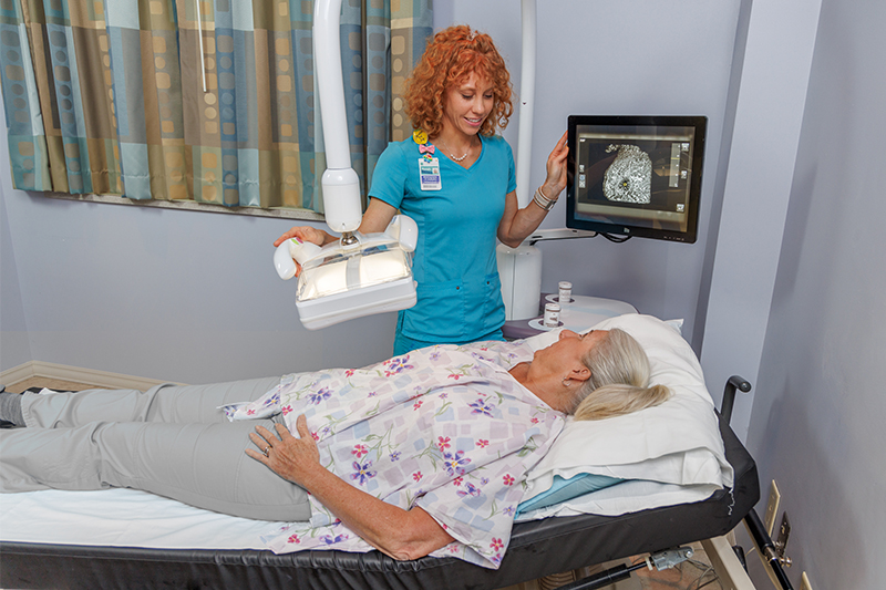
Beaufort Memorial offers an ultrasound technology that images both breasts at once. Known as automated breast ultrasound (ABUS) screening, this advanced approach captures hundreds of images in a short amount of time. Any areas of concern can then undergo further testing.
Like all breast imaging technology, ABUS protects your physical health, reducing mental stress and the need for unnecessary follow-up care.
“Our screenings provide a clearer view of the breast,” says Dr. Tara Grahovac, a board-certified, fellowship-trained breast surgical oncologist who specializes in the surgical management of breast disease at Beaufort Memorial. “This improves breast cancer detection and reduces the number of false positives that lead to unnecessary biopsies.”
Schedule your mammogram today by calling 843-522-5015, or request an appointment online.


